Most women who have had a Pap smear describe the same experience: a few uncomfortable minutes in a clinic, a swab, and then two weeks of waiting. What happens in between is almost entirely invisible.
That invisibility matters more than it sounds. In India, screening coverage remains strikingly low — one 2025 analysis of national data found that only about 1.9% of women aged 30 to 49 had been screened, and India accounts for a large majority of the cervical cancer burden across the WHO South-East Asia region. Globally, the International Agency for Research on Cancer estimated roughly 660,000 new cervical cancer diagnoses and 350,000 deaths in 2022.
Cervical cancer is one of the few cancers that can be caught and treated before it ever becomes cancer. The Pap smear is how. Understanding what the laboratory actually does with your sample makes the test less abstract — and makes it easier to know when a result deserves a follow-up question.
What a Pap Smear Actually Collects
A Pap smear does not test for cancer directly. It collects cells shed from the surface of the cervix, specifically from the transformation zone — the area where the smooth lining of the cervical canal meets the tougher lining of the outer cervix.
This zone matters because it is where almost all cervical precancers begin. Cells here are actively dividing and remodelling, which makes them more vulnerable to persistent infection with high-risk human papillomavirus (HPV).
The clinician uses a spatula, a brush, or a combined device to sample this area. What they collect is a population of cells, not a tissue structure. Everything that follows is an exercise in preserving those cells well enough that someone can look at them individually under a microscope.
The Six Stages Your Sample Goes Through
1. Collection and Preservation
There are two common approaches.
In a conventional Pap smear, the collected cells are spread directly onto a glass slide at the clinic and fixed immediately — usually with 95% ethanol or a spray fixative.
In liquid-based cytology (LBC), the collection device is rinsed into a vial of preservative fluid. The laboratory later processes that fluid to deposit a thin, even layer of cells onto a slide.
LBC has become more common because it reduces obscuring material like blood and mucus, and the residual fluid can often be used for HPV testing without a second visit.
2. Fixation — The Step That Decides Everything
This is the most underappreciated stage in the entire process.
Cervical cells begin to dry within seconds of being spread on a slide. Once they air-dry, the nuclei flatten and lose the fine chromatin detail that a screener relies on to distinguish a normal cell from a precancerous one.
Air-drying artifact is one of the most common reasons a conventional smear is judged unsatisfactory. It is not a laboratory failure — it happens in the clinic, in the seconds between spreading and fixing.
3. Staining
Cells are colourless. The Papanicolaou stain, developed by George Papanicolaou in the 1940s and still in use, is a polychromatic method: haematoxylin colours the nuclei, while orange G and eosin azure counterstains colour the cytoplasm in shades that vary with the cell's maturity and keratin content.
The result is diagnostically useful colour. A mature superficial squamous cell stains pink; a less mature one stains blue-green. Deviations from the expected pattern are part of what flags an abnormality.
4. Coverslipping
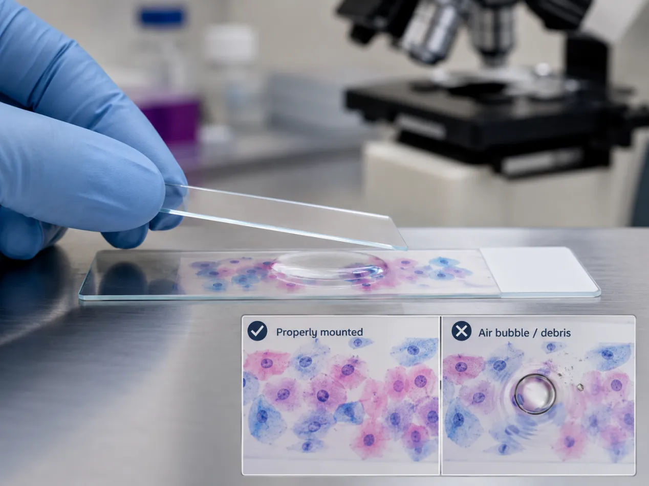
After staining, the slide is sealed with mounting medium and a thin sheet of glass placed over the cells.
This step is more consequential than it appears. Microscope objectives used in cytology are optically corrected for a specific glass thickness — typically around 0.17 mm — and the microscope slides and cover glass used in diagnostic laboratories are manufactured to tight dimensional and optical tolerances for exactly this reason. Glass that is off-specification, uneven, or not properly cleaned introduces optical distortion and can interfere with cell adhesion during processing.
Trapped air bubbles, incomplete coverage, or debris on the glass can obscure the very cells the screener needs to evaluate. In a test whose entire diagnostic value rests on resolving detail inside a single nucleus, these are not trivial variables.
5. Screening
A trained cytotechnologist examines the slide systematically — typically field by field, covering the entire cellular area.
This is genuinely difficult work. A single slide may contain tens of thousands of cells, and abnormal cells can be rare, scattered, and subtle. Many laboratories now use computer-assisted imaging to flag fields of interest, but human review remains central.
6. Pathologist Review
Slides screened as abnormal — and a random sample of those screened as normal, as a quality check — go to a pathologist for final interpretation and reporting.
Why Some Samples Come Back "Unsatisfactory"
An unsatisfactory result is not a diagnosis. It means the sample could not be reliably evaluated. Common causes include:
-
Insufficient cellularity. Adequacy standards require a minimum number of well-preserved squamous cells — substantially more for a conventional smear than for LBC.
-
Obscuring blood or inflammation. If more than a threshold proportion of cells are hidden, the slide cannot be read.
-
Air-drying artifact. As described above.
-
Absent transformation zone component. This is reported separately and does not always make a sample unsatisfactory, but it is noted because it affects how confident the negative result is.
If your report says unsatisfactory, the standard response is a repeat test, usually after a short interval. It is not a cause for alarm, but it is a reason not to simply file the report away.
Reading the Report: The Bethesda System
Most laboratories report Pap results using the Bethesda System, which uses standardised terminology rather than the older numbered classes. The main categories you may encounter:
|
Result |
What it means |
|
NILM |
Negative for intraepithelial lesion or malignancy — no abnormal cells found |
|
ASC-US |
Atypical squamous cells of undetermined significance — mildly abnormal, cause unclear |
|
ASC-H |
Atypical squamous cells, cannot exclude a high-grade lesion |
|
LSIL |
Low-grade squamous intraepithelial lesion — usually reflects active HPV infection |
|
HSIL |
High-grade squamous intraepithelial lesion — a genuine precancerous change |
|
AGC |
Atypical glandular cells — requires investigation, as it can originate above the cervix |
Two things are worth internalising. First, none of these are cancer diagnoses. LSIL in particular is common and frequently resolves on its own, especially in younger women. Second, HSIL is not cancer either — it is precancer, and it is treatable, which is precisely the point of screening.
What HPV Testing Adds
Since virtually all cervical cancer is caused by persistent high-risk HPV infection, testing for the virus directly is a more sensitive screening approach than looking for the cellular changes it eventually causes.
The World Health Organization now recommends HPV DNA testing as the preferred primary screening method for the general population, rather than cytology or visual inspection. In practice, many settings use HPV testing and cytology together, or use cytology to triage HPV-positive results.
If you are being screened, it is reasonable to ask which test you are receiving. They answer different questions.
Five Questions Worth Asking
-
Which test am I having — conventional cytology, liquid-based cytology, HPV testing, or a combination?
-
How was my sample fixed? For a conventional smear, immediate fixation matters.
-
Was my sample satisfactory for evaluation? This is stated on every report.
-
Was a transformation zone component present?
-
When should I be screened again, given my age and this result?
The Bottom Line
A Pap smear is a chain of small technical steps, and the result is only as reliable as the weakest link — how the sample was collected, how quickly it was fixed, how well it was stained and mounted, and how carefully it was screened.
None of this is a reason for anxiety. It is a reason to treat screening as something you participate in rather than something that happens to you. In a country where fewer than one in fifty eligible women is currently being screened, showing up at all is the single most important variable.
This article is for general information and is not a substitute for medical advice. Speak with a qualified healthcare provider about screening decisions appropriate to your age, history, and risk factors.

Reviewed by